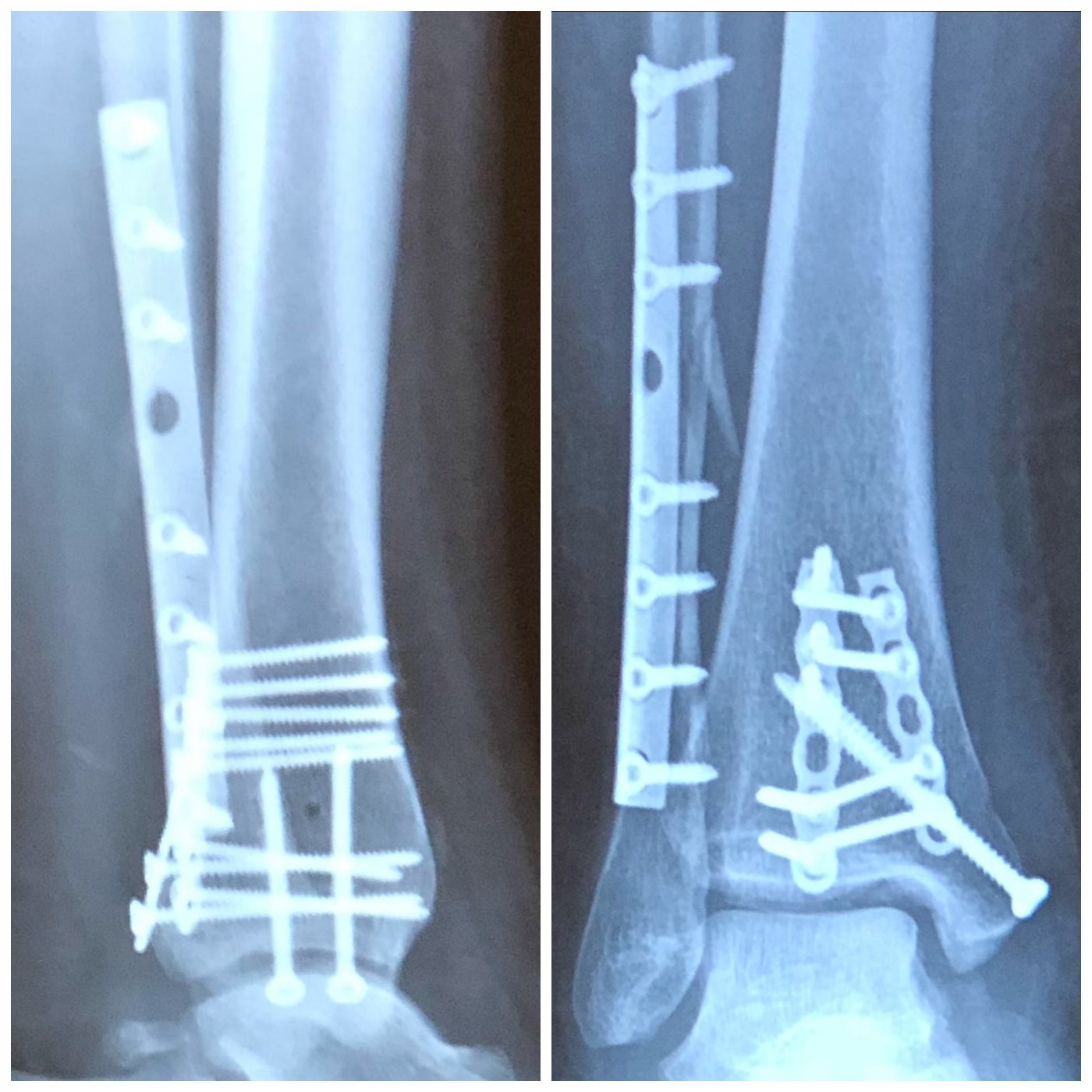
#Trimalleolar fx full
Many specialists prefer weight-bearing views, which may be very useful however, patients with severe foot and ankle injuries may have problems weight-bearing.The operative goal in the treatment of ankle fractures is well established: to restore anatomy and stability for early movement and full functional recovery, and prevent post-traumatic arthritis. In addition, a malunion in the anatomical relationship may indicate an old fracture (it is also necessary to look for bony sclerosis). An increase in the space between the talus and the surrounding fibula or tibia 5 mm or more is suspicious for a complete disruption of the lateral or medial collateral ligament. These views place an anatomical load on the ankle mortise. Stress views: usually obtained by specialists.Osteochondritis dissecans should be suspected in any patient who complains of joint locking or joint mice.
:max_bytes(150000):strip_icc()/bimalleolar-ankle-fractures-2549416_final-f8eed61efaec4254a6a60e4d5fae0c09.gif)
This latter condition appears as a minute semioval (saucer-shaped) bite mark along the joint line. Uses include detection of pilon fractures of the distal tibia or fractures of the talar dome as well as osteochondritis dissecans of the tibiotalar joint.
#Trimalleolar fx series
It may be used as part of acute trauma series for high-impact injuries. A mortise view eliminates the overlap of the distal fibula and tibia seen on routine AP projections and allows for better visualization of the talar dome and the distal tibia (plafond).


The mechanism of injury is direct impact caused by inversion or eversion injuries. Patients complain of marked pain, swelling, and tenderness associated with an inability to bear weight or ambulate. About one-third of all ankle fractures are bimalleolar and trimalleolar fractures.īimalleolar and trimalleolar fractures account for more than 30% of all ankle fractures.Trimalleolar fractures are fractures of the distal fibula, tibia, and the posterior lip of the distal tibia.Bimalleolar fractures are fractures of the distal fibula and tibia.


 0 kommentar(er)
